Tetragonal scheelite-like Ln:NaT(XO4)2 (T=Y, La, Gd, Lu; X= W, Mo) double tungstates and molybdates
Morphology-controlled trivalent lanthanide-doped Ln:NaGd(WO4)2 crystalline particles of the tetragonal scheelite-like structure phase were initially prepared through mild (170-200 ºC and autogenous pressure) hydrothermal (HT) syntheses with pH=6.0-7.5. Reaction times shorter than 8 h yield basically rod-like morphologies, although some quasi-spherical nanoparticles of ~5-20 nm can be also distinguished, see Figs. 1a-f. Prolonging the reaction time (10-12 h), nanosized octahedra appear, which coexist with nanorods, and finally longer reaction times (up 12 h) produce well-defined micron-sized octahedra, see Figs. 2a-e. The spectroscopic properties of Yb3+ in micron-sized HT NaGd(WO4)2 particles have been found to be equivalent to those obtained in bulk single crystals (i.e., Yb3+ single exponential photoluminescence decay and 2F5/2 lifetime of 330 µs), https://doi.org/10.1021/cm9032622.
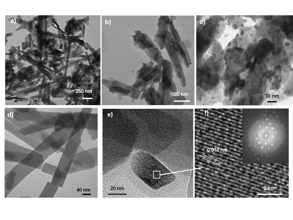 Figs. 1a-f. TEM and HRTEM images of Yb:NaGd(WO4)2 prepared by HT synthesis at 170 ºC and pH=6, with reaction times of 6 h (a, b) and 8 h (c-f)
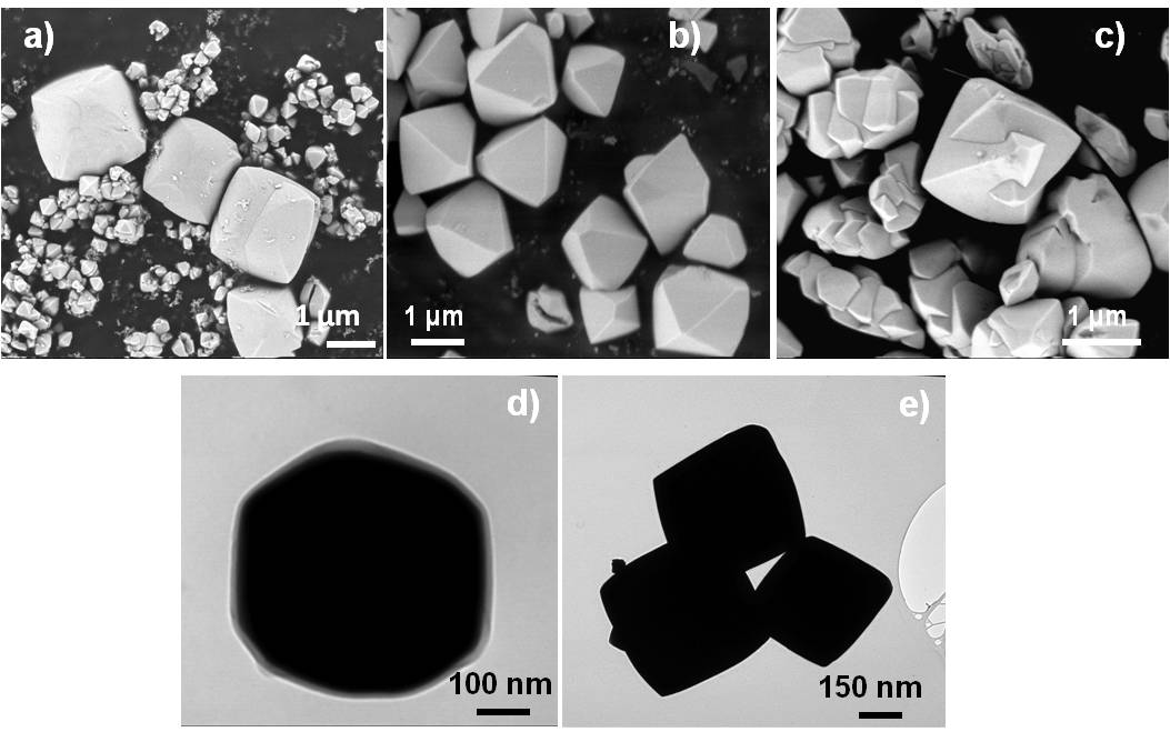 Figs. 2a-e. Images of Yb:NaGd(WO4)2 particles prepared by HT synthesis at 170 ºC and pH=7.5: FE-SEM micrographs for reaction times of 8 h (a), 12 h (b) and 14 h (c); TEM views of smaller octahedra obtained after reactions of 8 h (d, e).
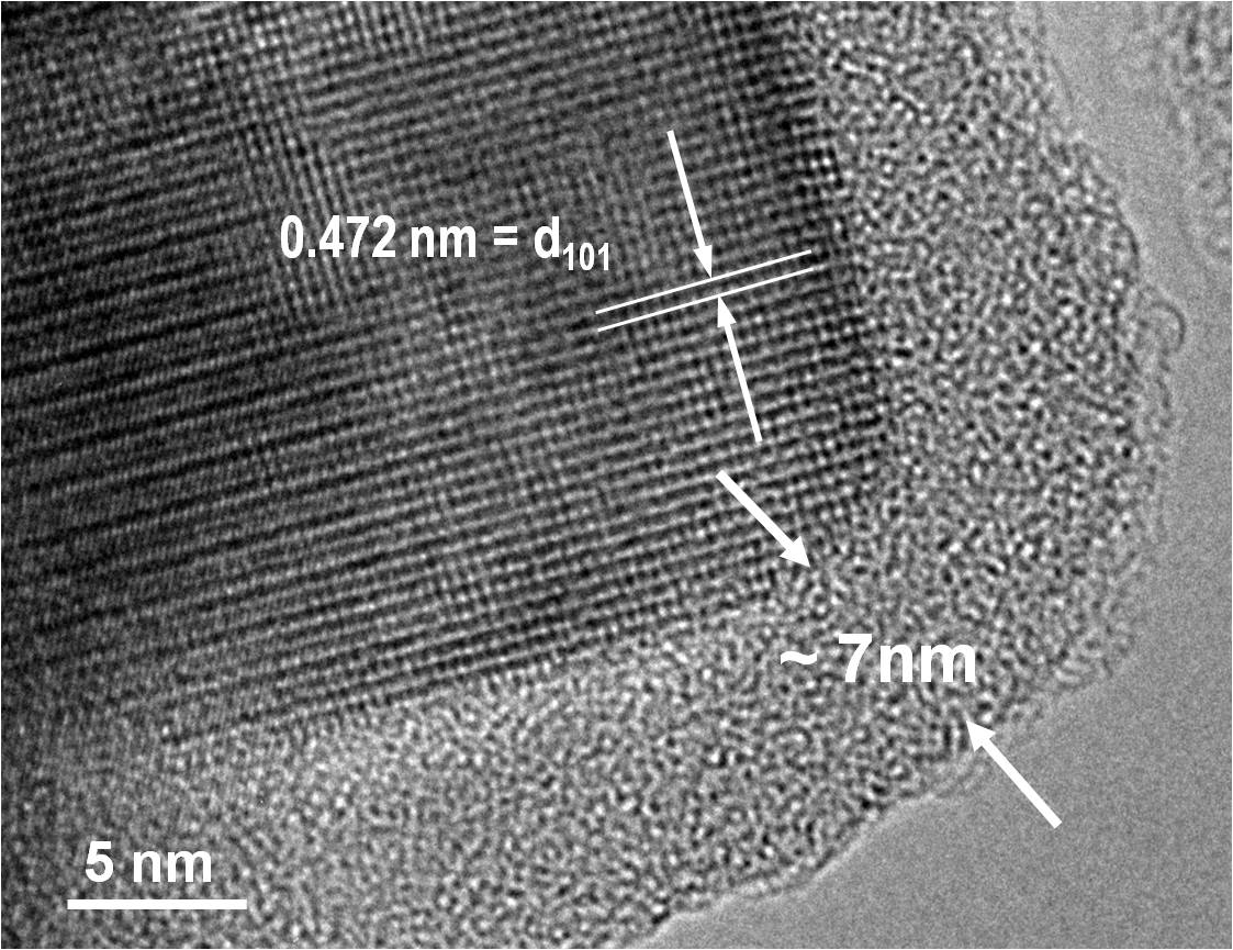
Coprecipitation syntheses in high boiling point solvents allowed obtaining ultrasmall diamond shaped Yb:Er:NaGd(WO4)2 crystalline nanoparticles (NPs) with diagonal dimensions in the 5–7 nm×10–12 nm range, Fig. 3, https://doi.org/10.1088/1361-6528/aa6834. The size of Yb:Er:NaGd(WO4)2 NPs can be selected by controlling the reaction conditions, and furthermore, these NPs have been coated with a shell of the isostructural compound NaY(MoO4)2, see the corresponding core-shell micrograph in Fig. 4.
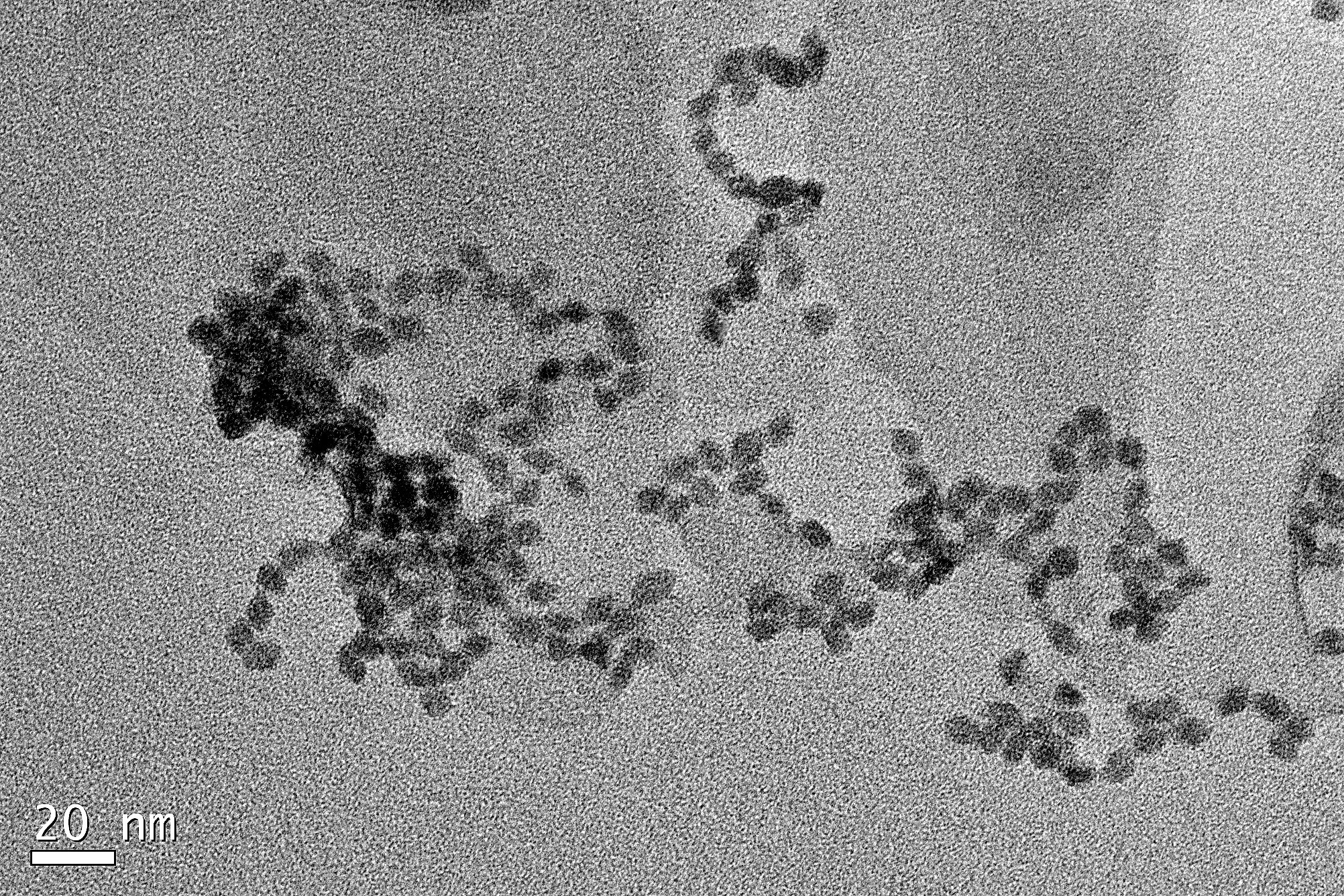
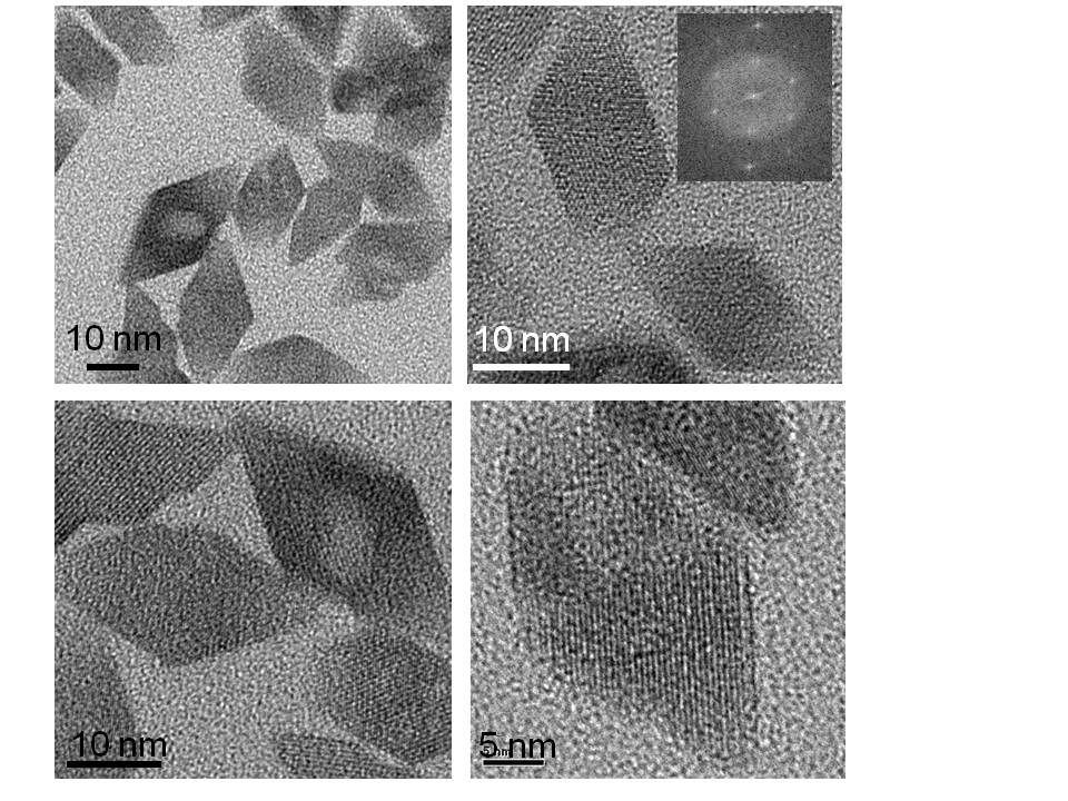 Fig 3. Diamond shaped Yb:Er:NaGd(WO4)2 crystalline NPs
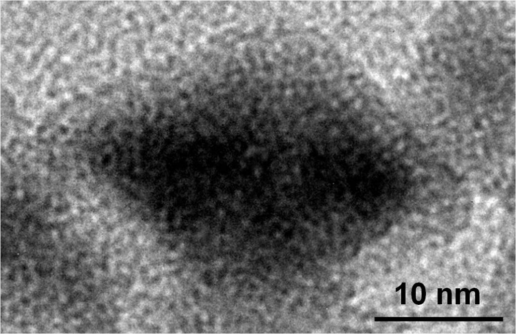 Fig. 4. Yb:Er:NaGd(WO4)2@NaY(MoO4)2 core-shell NPs
Our group has recently demonstrated that tetragonal scheelite-like Yb:Er:NaT(XO4)2 (T=Y, La, Gd, Lu, and X= Mo, W) compounds combine green UC efficiency comparable to that of Yb:Er:b NaYF4 (i.e. with quantum yield -QY- up to a quarter of that found for the latter), in spite of the large cutoff phonon energy of these oxides (~920 cm-1, about twice of that corresponding to the fluoride), and interestingly, thermal sensitivity S 2.5-5 times larger than that of the fluoride, at the temperature range of biological interest (~303-317 K/30-44ºC) S=108-118×10-4 K-1, being the largest values so far reported using the green Er3+ intensity ratiometric ratio (https://doi.org/10.1088/1361-6528/aa6834, https://doi.org/10.1371/journal.pone.0177596). In our experiments,cultured human mesenchymal stem cells (hMSCs) incubated with water emulsions of ultrasmall (diagonal dimensions 5-7 nmx10-12 nm) NPs of Yb:Er:NaGd(WO4)2 modified with TWEEN80 internalize and accumulate the NPs in endosomes/lysosomes, see Fig. 5, without degradation of the metabolic activity after 72 h of incubation (10 µg.ml-1 of NPs in the culture medium).
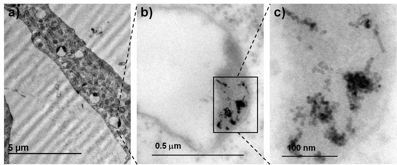 Fig.5 (a)–(c) Increasing magnification TEM images of hMSCs cultured for 24 h in a TWEEN80–modified 15 at%Yb:1 at%Er:NaGd(WO4)2 NP suspension (2.5 μg ml−1). NP accumulation is found embedded in a membrane-enclosed matrix, as lysosomes or late endosomes
The similar efficiency of Yb:Er:NaT(XO4)2 and Yb:Er: b-NaYF4 has allowed us testing equal subcutaneous depths (in excess of 2 mm) of ex-vivo chicken tissue in both cases, see the comparison in Fig. 6. Additionally, the UC signal of individual quasi-spherical (50-80 nm range) Yb:Er:NaLu(MoO4)2 NPs perfused in a mouse has been unequivocally identified by using fluorescence lifetime imaging microscopy (FLIM) and further compared with the SHG or autofluorescence images of the surrounding tissues, showing that these NPs reach all mouse organs, including heart, lung, kidney, liver, spleen, eye and brain, see Fig. 7a-f.
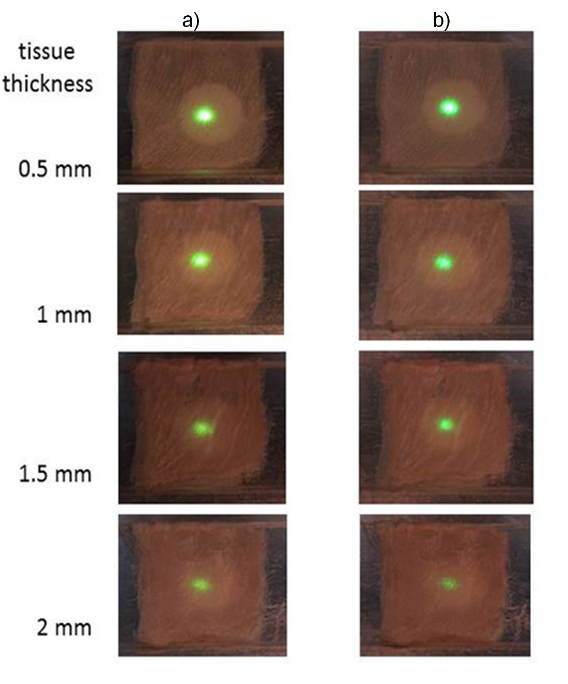 Fig. 6. Simulation of subcutaneous up-conversion detection. Images of the UC emission of the reference 7.5at%Yb:0.5%Er:β-NaYF4 (a) and of 15%Yb:1%Er:NaLu(MoO4)2 (b) compounds lying below several tissue thicknesses of ex-vivo chicken breast.
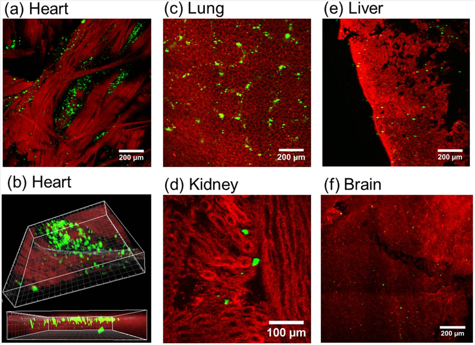
Fig. 7. Bi- and three dimensional micrographs of Yb:Er:NaLu(MoO4)2 NPs perfused in mouse organs, showing tissue autofluorescence (red, a, b, d, e, f), SHG (c) and UC (green).
Orthorhombic Pbcm Vernier-type Ln-doped Y6O5F8 oxyfluorides
Micron-sized bundles of highly crystalline individual nanotubes of Yb3+-sensitized, Ln3+(Er3+, Pr3+)-doped Y6O5F8 oxyfluorides (orthorhombic Pbcm Vernier-type structure) have been prepared through efficient hydrothermal synthesis at 185ºC, (https://doi.org/10.1039/C4CP03616F), see Figs. 8a-d.
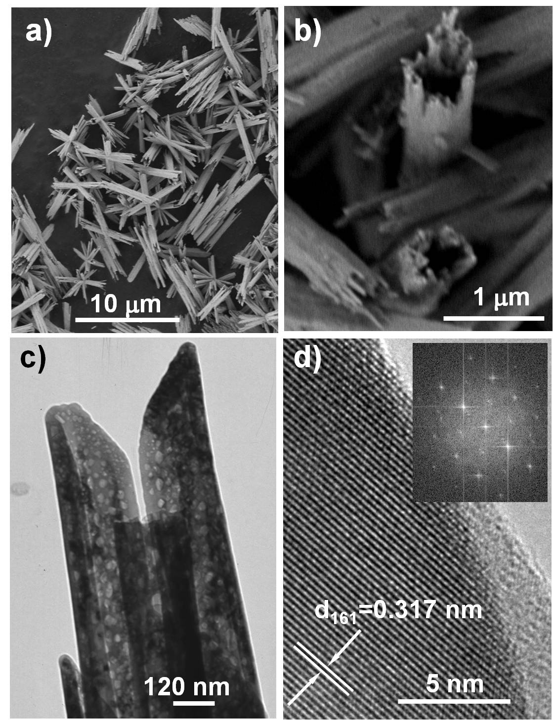 Fig. 8. Images of Er, Yb:Y6O5F8 : (a) and (b) SEM images of micron-sized bundles. (c) TEM image of nanotubes forming bundles. (d) HRTEM image of a nanotube and its corresponding Fast Fourier Transform (FFT) image in the inset.
The ratiometric analysis of the thermal evolution of intensities of Er,Yb:Y6O5F8 NIR ( ∼978 nm)-excited green upconverted emissions from thermally coupled 2H11/2 and 4S3/2 Er3+ multiplets indicates thermal sensing potential with very high sensitivity S = 0.0060 K-1 at physiological temperatures, which surpasses the S value found for Er, Yb:β-NaYF4 at these temperatures.
Also under NIR diode laser excitation, the color of the upconverted light from codoped Pr, Er, Yb:Y6O5F8 nanotubes can be selected by the control of the Pr3+ concentration and by the excitation regime and power density. Samples with low Pr3+ concentration emit green light, and the selection between bluish-green light and white light has been demonstrated with high Pr3+ concentration (2 mol%), under pulsed or continuous wave excitation, respectively, see Fig. 9a-c.
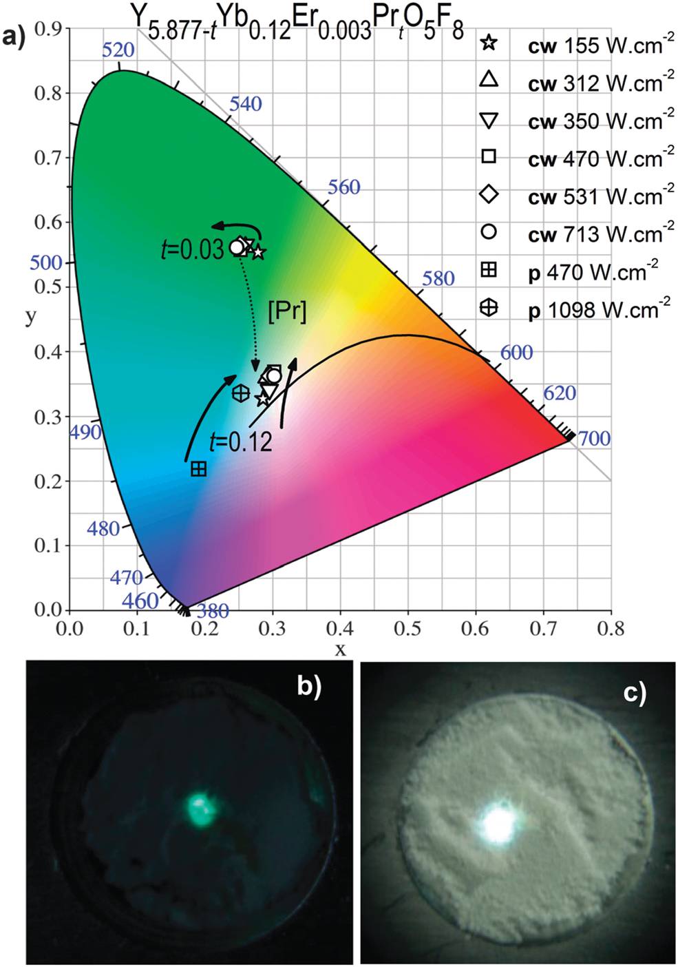
Fig. 9. (a) Variation of the CIE coordinates for Y5.87-tYb0.12Er0.003PrtO5F8 with t = 0.03 (0.5 mol%) and t = 0.12 (2.0 mol%) under NIR cw excitation at 978 nm. The black line corresponds to the black-body radiation; (b) and (c) Digital images of the bluish-green light emission from the sample with 0.5 mol% Pr (cw excitation of 784 W cm-2 power density) and of UC white light from the sample with 2.0 mol% Pr (pulsed excitation of 627 W cm-2 power density), respectively.
Tetragonal zircon-type Ln:GdVO4 vanadates
Chemical processes involving low temperature hydrothermal treatments of pH 4, 7 and 10 solutions of Gd(Tm)-nitrates or chloride reagents and NH4VO3 result in the formation of crystalline nanorods, nanotubes, nanoribbons, nanospindles or 3D micro- and nanoparticles of lanthanide (Ln)-doped tetragonal zircon-type GdVO4 vanadates. Each of these morphologies are critically depending on the structural arrangement generated by specific V5+-molecular precursors as well as the hydrothermal conditions. (https://doi.org/10.1039/c2ce06515k).
Codoped Yb,Ln:GdVO4 (Ln=Tm, Ho, Er) upconverting crystalline nanoparticles (NPs) with square and rectangular sections (side lengths of 25–35 nm) were prepared through hydrothermal synthesis at pH 7 and 185ºC during 24 h. Further coating of the surface with a uniform 5 nm-shell of SiO2 results in a significant improvement of the intensity of the upconverted emitted visible light following near-infrared (lEXC~980 nm) diode laser excitation with respect to raw hydrothermal NPs. These processed samples yield bright visible light, which is composed of red (predominant)-green, blue and green emissions from Ho3+, Tm3+ and Er3+, respectively. Based on analysis of the upconversion mechanisms and from calculations of the CIE color coordinates, the nature of the doping Ln and its concentrations as well as the applied DL excitation power densities have been determined to obtain any color or for bright white light generation. Nearly ideal white upconverted light has been achieved for samples of composition Gd0:829Yb0:15Tm0:01Ho0:009Er0:002VO4. (https://doi.org/10.1088/0957-4484/23/50/505205. Article Selected as Nanotechnology Publishers´s pick of December 2012).
Furthermore, Er,Yb:GdVO4@SiO2 core−shell NPs of the above described morphology can be used as an efficient luminescence temperature sensor based on ratiometric upconversion measurements. We demonstrate the benefits of the SiO2 coating on the luminescent thermometric properties. By using a SiO2 coating, a ×2 enhancement of the thermal sensitivity was achieved. Additionally, the SiO2 shell protects the NPs from overheating during the excitation process. Also, we demonstrated that a high thermal resolution can be achieved using these core−shell NPs. The spatial resolution is estimated to be of the order of the size of the core−shell NPs. Good stability in different solvents, such as water, methanol, and DMSO, make it possible to use them as a temperature sensor in biological applications, as in the one we reported to monitor an induced heating process in an ex vivo experiment (https://pubs.acs.org/doi/10.1021/acsami.6b01371).
We also propose the first, to the best of our knowledge, thermochromic temperature small scale sensor based on the blue to deep red color change of the upconverted light from Yb; Tm: GdVO4@SiO2 core–shell NPs under ~980 nm excitation. The electronic coupling of the 1G4 (blue) and 3F2 (deep red) Tm3+ emitting levels has been experimentally evidenced. Energy transfer schemes accounting for the thermal evolution of the electronic populations of these energy levels have been proposed to support the temperature dependent ratiometric relationship between the intensities of the visible signals, thus providing the internal calibration to the temperature sensing system. The potentiality of this nanothermometer was demonstrated by monitoring the heating process produced by the Joule effect in a Pt wire of 50 µm in diameter, being the thermal and temporal resolutions ±0.1 K and <16 ms, respectively. The results matched very well with the theoretical modeling for this system (https://doi.org/10.1039/c6tc01841f).
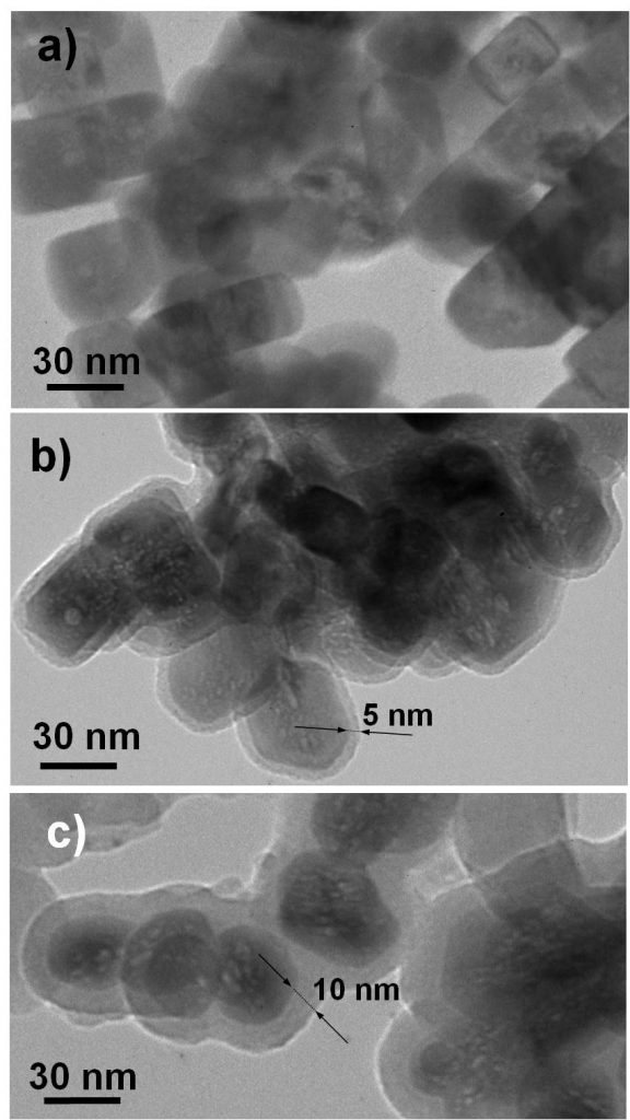 TEM images of nanocrystalline Yb, Tm-GdVO4 samples: a) NPs with square and rectangular sections of the bare hydrothermal (24h at 185 ºC, pH 7) prepared sample, b) NPs coated with a 5 nm-shell of SiO2 after reaction with TEOS, c) NPs coated with a 10 nm-shell of SiO2.
 HRTEM image of one Yb, Er:GdVO4@SiO2 NP