INSTRUMENTATION

Experimental Probes and Facilities

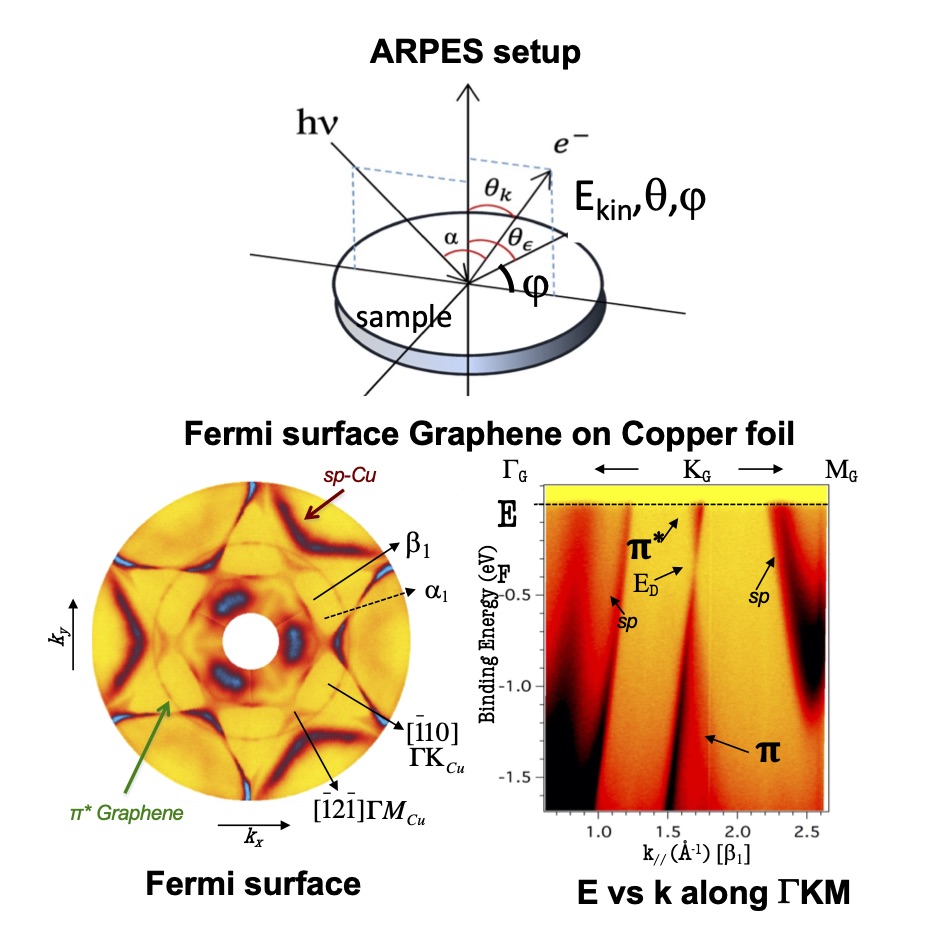
  • Angle-Resolved Photoemission Spectroscopy, (ARPES)

ARPES is an experimental technique that directly measures the binding energy of the emitted electrons’ initial state as a function of the reciprocal space’s momentum photons of a well-defined energy is incident upon a simple. ARPES enables direct observation of the Fermi surface and the underlying electronic structure of crystals, which are fundamental attributes of any material that allows describing all the electronic properties of solids and reveal the nature of vital electronic interactions involved. ARPES has proved to be particularly fruitful in studying quasi-1D and -2D materials, where the photoemission no conservation of the perpendicular k vector to the surface is not affecting the measurements. Traditionally, angle-resolved photoemission spectroscopy (ARPES) is the only technique capable of making sufficiently precise measurements of the dispersion of the band structure of materials in the reciprocal space. The state of the art ARPES equipment installed at synchrotron radiation sources is such that it can offer energy and angular resolution of better than 5 meV and 0.1Å, respectively. Yet, until now, no instrument has been capable of performing spatially resolved ARPES experiments on the nanometer scale.

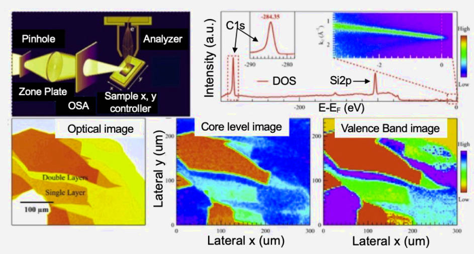
Our group has designed, built, installed, commissioned, and launched the standard operation of a Nano-ARPES (Nano Angle Resolved Photoelectron Spectroscopy) beamline named ANTARES at the SOLEIL synchrotron in France. This sophisticated instrument is able, with a spatial resolution of several tens of nanometers, of carrying out the direct imaging of core levels, their chemical shifts, band electronic structures, and constant energy surfaces in the reciprocal space, especially the Fermi surfaces. This cutting-edge technique also named k-space nanoscope is an innovative and powerful tool able to nano-imaging the electronic structure, chemistry, and functional composition of non-homogeneous samples as well as very tiny materials of the order of nano- and mesoscopic-scale samples.

The k-nanoscope is a spectroscopic non-destructive nano-probe to study advanced materials. This innovative scanning photoemission nanoscopy combines linear and angle sweeps to perform the electronic and chemical imaging of tiny and heterogeneous samples can be precisely performed by detecting the electronic band structure, the Fermi surface, and the chemical functional composition point by point throughout the surface of the whole sample, using the same k-nanoscope setup. The design effectively integrates insertion devices together with high photon tuning and transmission optics. Moreover, the photon source has been combined with advanced mechanical nano-positioning motors that ensure precise and reproducible sample handling. The setup working in ultra-high vacuum condition is fully compatible with a high angular and energy-resolved R4000 Scienta hemispherical analyzer and a set of Fresnel Zone Plates able to focalize the beam spot up to a few tens of nanometers.

Also, in the context of the MATINÉE, CSIC Research Unit, between Valencia University and the ICMM, our group collaborates in keeping operative a brand-new ARPES and Spin-ARPES setup installed at the Institute of Materials Sciences of Valencia University (ICMUV).  

ARPES SETUP, FERMI SURFACE AND BAND DISPERSION

NANO-ARPES SETUP, CHEMICAL AND ELECTRONIC IMAGING

k-SPACE NANOSCOPE

  • Synchrotron and Neutron facilities: 

          Neutron sources

Neutrons are unique as probes into the matter; they can reveal what other techniques cannot detect. They, therefore, provide a powerful tool for investigating the intimate structure of materials, playing a crucial role in modern scientific research. Our group is a regular user of the Institut Laue–Langevin (ILL) in Grenoble, France. Mainly neutron diffraction experiments of mono and poly-crystals samples are carried out using several experimental stations of the ILL.

           Synchrotron Radiation Sources

For high-resolution synchrotron ARPES and core-level photoemission and X-ray absorption like NEXAFS, Synchrotron Radiation sources offer flexibility in varying photon energy, optimizing matrix element effects to optimize ARPES spectra and tune high and low core level edges for NEXAFS. Our group uses several European Large Facilities, frequently working in different beamlines, depending on the type of the samples to investigate. Mainly, we carried out ARPES, nano-ARPES, core-level photoemission, and NEXAFS experiments in the photon energy range of 21 eV to 1000 eV in ANTARES beamline in SOLEIL, ELETTRA, MAX IV, ESRF, ALS, and ALBA synchrotron sources.

European Neutron Sources

Synchrotron Radiation Facilities

European Synchrotron Radiation Facility

  • High pressure laboratory – High Hydrostatic pressure

The high pressure equipment we set up in our laboratory allows us to explore a wide working field that, until now, was restricted to a few groups, given the scarce availability of the heavy instrumentation which was, traditionally, associated with the use of high pressure (belt-type or multi-anvil type presses, with working pressures of 4-10 GPa)


  • Fuel Cells

The preparation of cathode materials for SOFC is carried out by citrate techniques, simply followed by a thermal treatment in air at the adequate temperature to stabilize the wanted phase. The preparation of anode materials requires, by contrast, the treatment of the previously “oxidized” sample into a “reduced” material, which must be stable in the reducing conditions of the fuel (H2 or CH4). 

  • Nuclear Magnetic Resonance (RMN) laboratory

Nuclear Magnetic Resonance (NMR) Laboratory expertise from Intertek supports clients across the world. NMR laboratory observation of any NMR-active nucleus in the periodic table is posible. NMR is a powerful technique that can provide information on molecular structure and dynamics at the atomic level. It has been widely used in chemistry, polymer and biology, with rapid analysis and reporting times. NMR active nuclei studied include 1H, 11B, 13C, 19F, 27Al, 29Si, and 31P.

NMR laboratory instrumentation:  500 MHz NMR. Z-gradients for diffusion measurements and gradient-selected techniques. Multidimensional techniques: 1H, correlation spectroscopy (1H-COSY), Total correlation spectroscopy (TOCSY), Heteronuclear single-quantum correlation spectroscopy (HSQC), Heteronuclear multiple-bond correlation spectroscopy (HMBC), Nuclear Overhauser effect spectroscopy (NOESY), and Incredible natural-abundance double-quantum transfer experiment (INADEQUATE).

  • Electrochemical Characterization of Batteries

Electrochemistry is a powerful tool that allows us to determine important properties of  propttyipical batteries.These analyses are performed in three-electrode cells, where the voltage against a reference electrode is measured while a current is applied between a counter and a working electrode.

Fully automated electrochemical setup

At the GREENER group, we have a fully automated electrochemical setup, which allow us to perform measurements on ten different samples simultaneously.  We have standardized protocols for activity and stability measurements of multiple electrochemical reactions. We can measure on particles, wires and polycrystalline surfaces, in both acid and alkaline media. Combined analysis methods give the best results.