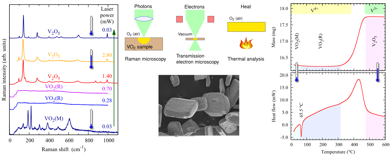
P. Vilanova-Martínez, J. Hernández-Velasco, A. R. Landa-Cánovas, and F. Agulló-Rueda, “Laser heating induced phase changes of VO2 crystals in air monitored by Raman spectroscopy,” J. Alloys Comp. 661, 122–125 (2016)
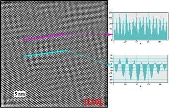
 HRTEM image of a crystal of oxidized ∼SbVO4 orientated along the [1 1 0] rutile direction. The white dots correspond to basic rutile cation positions and the superimposed dark bands to sinusoidal waves of cation vacancies. The magenta and blue lines indicate the line scans whose intensity is displayed at the right of the figure. Magenta line runs across the cationic positions while the blue line runs mainly across the cation vacancy waves. A. R. Landa-Cánovas, F. J. García-García y S. Hansen. «Structural Flexibility in ~SbVO4» Catalysis Today 158, 156–161 (2010).
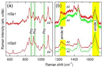
 Raman spectra of bioinspired silk fibers spun from solutions of two recombinant spidroin-like proteins [M. Elices et al, Macromolecules 44(5), 1166-1176 (2011)].
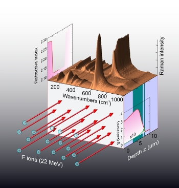
 Raman spectra as a function of depth for a LiNbO3 sample after irradiation with F ions at 22 MeV and a fluence of 3 × 10^15 cm–2 [A. Rivera et al, Phys. Status Solidi A 206, 1109–1116 (2009).]

