Study of the formation mechanism of hierarchical silicon structures produced by sequential ion beam irradiation and anodic etching

In a collaboration with the Autonomous University of Madrid (Department of Applied Physics and the Center for Micro Analysis of Materials) and the Institut für Solarenergieforschung Hameln (ISFH) of Germany we have studied the formation of micropatterns combining nanostructured (porous) Si (NPSi) and bulk Si by a sequential process of selective high energy ion irradiation and anodic etching.

E. Punzón Quijorna, S. Kajari-Shrönder, F. Agulló-Rueda, M. Manso Silván, R. J. Martín Palma, P. Herrero Fernández, V. Torres-Costa, and A. Climent, “Study of the formation mechanism of hierarchical silicon structures produced by sequential ion beam irradiation and anodic etching,” Vacuum 138, 238–243 (2017).

Publicado en HRTEM, ion irradiation, microscopy, nanocharacterization, nanoscience, nanotechnology, Raman spectroscopy, silicon | Etiquetado , , , , , , | Comentarios desactivados en Study of the formation mechanism of hierarchical silicon structures produced by sequential ion beam irradiation and anodic etching

Direct laser writing of nanorough cell microbarriers

In collaboration with the Advanced Photonic and Biofunctional Materials group the Autonomous University of Madrid, lead by Dr. Miguel Manso, we have studied by Raman microspectroscopy laser written silicon-based structures for biological cell microbarriers.

Y. Xiang, R. Martínez Martínez, V. Torres-Costa, F. Agulló-Rueda, J. P. Garcia-Ruiz, and M. Manso Silván, «Direct laser writing of nanorough cell microbarriers on anatase/Si and graphite/Si,» Mater. Sci. Eng. C 66, 8–15 (2016)

Publicado en microscopy, nanocharacterization, nanoscience, nanotechnology, Publications, Raman spectroscopy, silicon | Etiquetado , , , , , | Deja un comentario

Variability of silkworm silk and degumming

In collaboration with the Department of Materials Science of the Polytechnical University of Madrid we have studied by Raman spectroscopy as spun silkworm (Bombyx mori) silk fibers subjected to different degumming treatments.

G. B. Perea, C. Solanas, N. Marí-Buyé, R. Madurga, F. Agulló-Rueda, A. Muinelo, C. Riekel, M. Burghammer, I. Jorge, J. Vázquez, G. R. Plaza, A. Torres, F. del Pozo, G. V. Guinea, M. Elices, J. L. Cenis, and J. Pérez-Rigueiro, “The apparent variability of silkworm (Bombyx mori) silk and its relationship with degumming,” Eur. Polym. J., 78, 129–140 (2016).

Publicado en microscopy, Publications, Raman spectroscopy, silk | Etiquetado , , , | Deja un comentario

Jon Canca Ruiz has joined our group

Jon Canca Ruiz graduated in Chemistry from the University of the Basque Country. He has a Master on New Materials from the same university. He has worked on the synthesis and x-ray diffraction characterization of hybrid polyoxomethalates/metalorganic single crystals.

Publicado en People | Deja un comentario

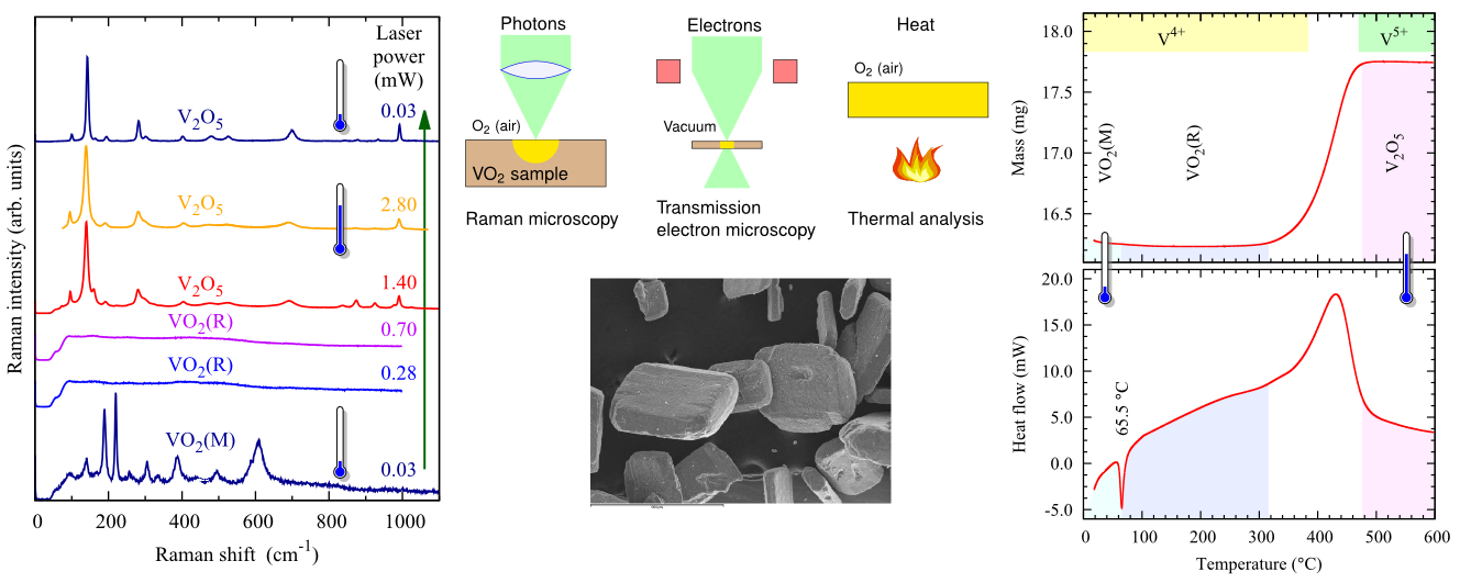
Laser heating induced phase changes of VO2

Vilanova2015GraphicalAbstract

P. Vilanova-Martínez, J. Hernández-Velasco, A. R. Landa-Cánovas, and F. Agulló-Rueda, “Laser heating induced phase changes of VO2 crystals in air monitored by Raman spectroscopy,” J. Alloys Comp. 661, 122–125 (2016)

Publicado en HRTEM, Publications, Raman spectroscopy, Sin categoría | Etiquetado , , , , , , , | Deja un comentario

HRTEM microscopy of transformations in bulk amorphous silica produced by ultrafast laser direct writing

A transmission electron microscopy study of nanogratings formed in bulk amorphous silica by direct writing with an ultrafast pulsed laser with a radiation wavelength of 1030 nm and pulse duration of 560 fs is presented. The results achieved show that the nanogratings are composed of planar nanostructures with an average periodicity of 250 nm and typical thickness of about 30 nm, consisting of alternating layers of heavily damaged material and layers of material where a dense precipitation of nanocrystals occurred. The crystallization of silica to form these nanocrystals can be explained by the large pressures and temperatures reached in these regions as a result of nanoplasma formation and recombination.

Collaboration with Instituto Superior de Engenharia de Lisboa, ICEMS—Instituto de Ciência e Engenharia de Materiais e Superfícies, and Instituto Superior Técnico, Lisbon, Portugal.

Vitor Oliveira, Sahendra P. Sharma, Pilar Herrero, and Rui Vilar, «Transformations induced in bulk amorphous silica by ultrafast laser direct writing,» Opt. Lett. 38, 4950 (2013).

Publicado en HRTEM, microscopy, nanocharacterization, nanoscience, nanotechnology, Sin categoría | Etiquetado , , , , | Deja un comentario

Chemical analysis of pigments in Velazquez paintings

The journal Vibrational Spectroscopy has published a work of us in collaboration with researchers Carolina Gutiérrez and Aurelio Climent, from the Autonomous University of Madrid (UAM), and Carmen Garrido, from the Prado Museum. In this work we have studied the chemical composition and the crystal structure of pigments in Diego Velazquez paintings from different periods in the artist life. Using Raman microspectroscopy we have found that the number of pigments was very limited and did not change significantly during the artist lifetime. P. C. Gutiérrez, F. Agulló-Rueda, A. Climent-Font, and C. Garrido, «Identification of pigments in Diego Velázquez paintings by Raman microscopy,» Vibrat. Spec. 69, 13–20 (2013)

La revista Vibrational Spectroscopy acaba de publicar un trabajo nuestro realizado en colaboración con los investigadores Carolina Gutiérrez y Aurelio Climent, de la Universidad Autónoma de Madrid (UAM), y Carmen Garrido, del Museo del Prado. En este trabajo hemos estudiado la composición química y la estructura cristalina de pigmentos en las pinturas de Diego Velazquez procedentes de distintos periodos en la vida del artista. Mediante microespectroscopía Raman hemos encontrado una paleta reducida de pigmentos que no cambió de forma significativa a lo largo de la vida del artista. P. C. Gutiérrez, F. Agulló-Rueda, A. Climent-Font, and C. Garrido, «Identification of pigments in Diego Velázquez paintings by Raman microscopy,» Vibrat. Spec. 69, 13–20 (2013)

Publicado en art, microscopy, Publications, Raman spectroscopy | Etiquetado , , , | Deja un comentario

Microscopy course starts on May 16th, 2013

The course Microscopy in Materiales Science: TEM and Raman 2013 is scheduled from May 16th to May 24th, 2013, at the Materials Science Institute of Madrid (ICMM-CSIC). The classes will be held in the afternoon. More details in the course webpage.El curso Microscopía en Ciencia de Materiales: TEM y Raman 2013 está programado del 16 al 24 de mayo de 2013. Las clases se impartirán por las tardes en el Instituto de Ciencia de Materiales de Madrid (ICMM-CSIC). Más información en la página web del curso.

Publicado en course, education, microscopy, Sin categoría, teaching | Deja un comentario

Microscopy course announcement

The course Microscopy in Materiales Science: TEM and Raman 2013 will start on May. The course will last two weeks and classes will be held in the afternoon. More details in the course webpage.El curso Curso de Microscopía en Ciencia de Materiales: TEM y Raman 2013 empezará en mayo. El curso dura dos semanas y las clases son por la tarde. Más detalles en la página web del curso.

Publicado en course, education, microscopy, teaching | Deja un comentario

Paloma Vilanova joins our group

Paloma VilanovaPaloma Vilanova-Martínez has joined our group. Paloma graduated in Chemistry from the Autonomous University of Madrid. She is working on the study of the micro- and nanostructural flexibility of mixed oxides with interest in catalysis (Project FlexOCat).

Paloma VilanovaPaloma Vilanova-Martínez se ha incorporado en nuestro grupo. Paloma se licenció en Química por la Universidad Autónoma de Madrid. Está trabajando en el estudio de la flexibilidad micro- y nanoestructural de óxidos mixtos con interés en catálisis (Proyecto FlexOCat).

Publicado en People | Deja un comentario